
-
 DE
DE



Chiari Malformation: Causes, Symptoms & Treatment
Chiari malformation is a structural brain defect in which part of the cerebellum extends into the spinal canal, potentially disrupting cerebrospinal fluid flow and causing neurological symptoms.
Add as Preferred SourceThings worth knowing about "Chiari malformation"
Chiari malformation is a structural brain defect in which part of the cerebellum extends into the spinal canal, potentially disrupting cerebrospinal fluid flow and causing neurological symptoms.
What Is Chiari Malformation?
Chiari malformation (also known as Arnold-Chiari malformation) is a structural abnormality of the brain in which parts of the cerebellum – specifically the cerebellar tonsils – protrude downward through the foramen magnum (the large opening at the base of the skull) into the upper spinal canal. This can obstruct the normal flow of cerebrospinal fluid (CSF), leading to a wide range of neurological symptoms. The condition was first described in the 19th century by Austrian pathologist Hans Chiari.
Types of Chiari Malformation
There are four main types:
- Type I: The most common type. The cerebellar tonsils extend more than 5 mm into the spinal canal. Symptoms often do not appear until adulthood. Frequently associated with syringomyelia (a fluid-filled cavity within the spinal cord).
- Type II: A more severe form that is almost always associated with myelomeningocele (open spina bifida). Presents in childhood.
- Type III: A rare and severe form involving herniation of the cerebellum and brainstem outside the skull.
- Type IV: A rare form characterized by incomplete development of the cerebellum (cerebellar hypoplasia).
Causes
The exact causes of Chiari malformation are not fully understood. It is believed that an abnormally small posterior cranial fossa (the part of the skull that houses the cerebellum) pushes brain tissue downward. Possible contributing factors include:
- Genetic changes and familial predisposition
- Disruptions during early fetal development
- Nutritional deficiencies during pregnancy (e.g., insufficient folate intake)
- Rarely: acquired forms following injury, infection, or persistently elevated CSF pressure
Symptoms
Many individuals with Type I remain asymptomatic for a long time. When symptoms occur, they may include:
- Headaches, particularly at the back of the head, often triggered by coughing, sneezing, or physical exertion
- Neck and shoulder pain
- Numbness, tingling, or weakness in the arms and hands
- Balance and coordination problems
- Difficulty swallowing or speaking
- Visual disturbances (e.g., double vision, blurred vision)
- Dizziness and tinnitus (ringing in the ears)
- In cases of syringomyelia: additional spinal cord symptoms such as loss of pain and temperature sensation
Diagnosis
The most important diagnostic tool is magnetic resonance imaging (MRI) of the head and cervical spine. MRI allows detailed assessment of the position of the cerebellum, brainstem, and CSF flow patterns. Phase-contrast MRI (PC-MRI) can be used to evaluate CSF flow through the foramen magnum. A computed tomography (CT) scan may be used as a complement to assess bony structures.
Treatment
Not all patients require treatment. In the absence of symptoms, a watchful waiting approach with regular follow-up imaging is often recommended.
Conservative Treatment
For mild symptoms, pain-relieving medications and physiotherapy may help manage discomfort.
Surgical Treatment
For significant or progressive symptoms, surgical decompression is the treatment of choice. The most common procedure is suboccipital decompression (also called foramen magnum decompression): a portion of the occipital bone is removed to create more space at the foramen magnum and restore normal CSF flow. This is often combined with a duraplasty, in which the dura mater (the tough outer membrane of the brain) is expanded using a patch. In many cases, an associated syringomyelia resolves or improves following decompression surgery.
Prognosis
The clinical course varies greatly between individuals. Many patients with Type I experience a stable condition without progression. After successful surgery, most patients report significant improvement in symptoms. Regular neurological follow-up and MRI monitoring are recommended.
References
- Greenberg, M.S.: Handbook of Neurosurgery, 9th edition. Thieme Medical Publishers, 2020.
- Meadows, J. et al.: "Chiari I malformations: radiological and morphometric analysis." Neurosurgery, 52(5): 1044–1052, 2003.
- National Institute of Neurological Disorders and Stroke (NINDS): Chiari Malformation Fact Sheet. National Institutes of Health (NIH), 2023. Available at: www.ninds.nih.gov

Building Blocks for a Healthy Life
As an integrated manufacturer, we develop & produce evidence-based, patented, and non-patented formulations. We work exclusively with plant and natural extracts according to the strictest purity & quality standards.
The combination of science & modern technology with the laws of nature creates solutions consistently tailored to humans – for the highest good: health.
Skip product gallery


Subscription available
Floral
For Healthy Oral Flora & Dental Care
Formulated lozenges with Dentalac®, lactic acid bacteria, and Lactoferrin CLN®30 lozenges
Regular price:
€22.90
Discount
Subscription available
Lactoferrin 60 Capsules

For your universal protection
As one of the most valuable proteins in the body, lactoferrin is a natural component of the immune system.
60 Capsules
Sale price:
€50.00
Regular price:
€59.90
Discount
Subscription available
Multivitamin Mineral Complex
Daily basic supply: the complete spectrum of micronutrients
Natural multivitamin complex for the immune system, energy, and central body functions
30 Capsules
Sale price:
€27.90
Regular price:
€29.90
Subscription available
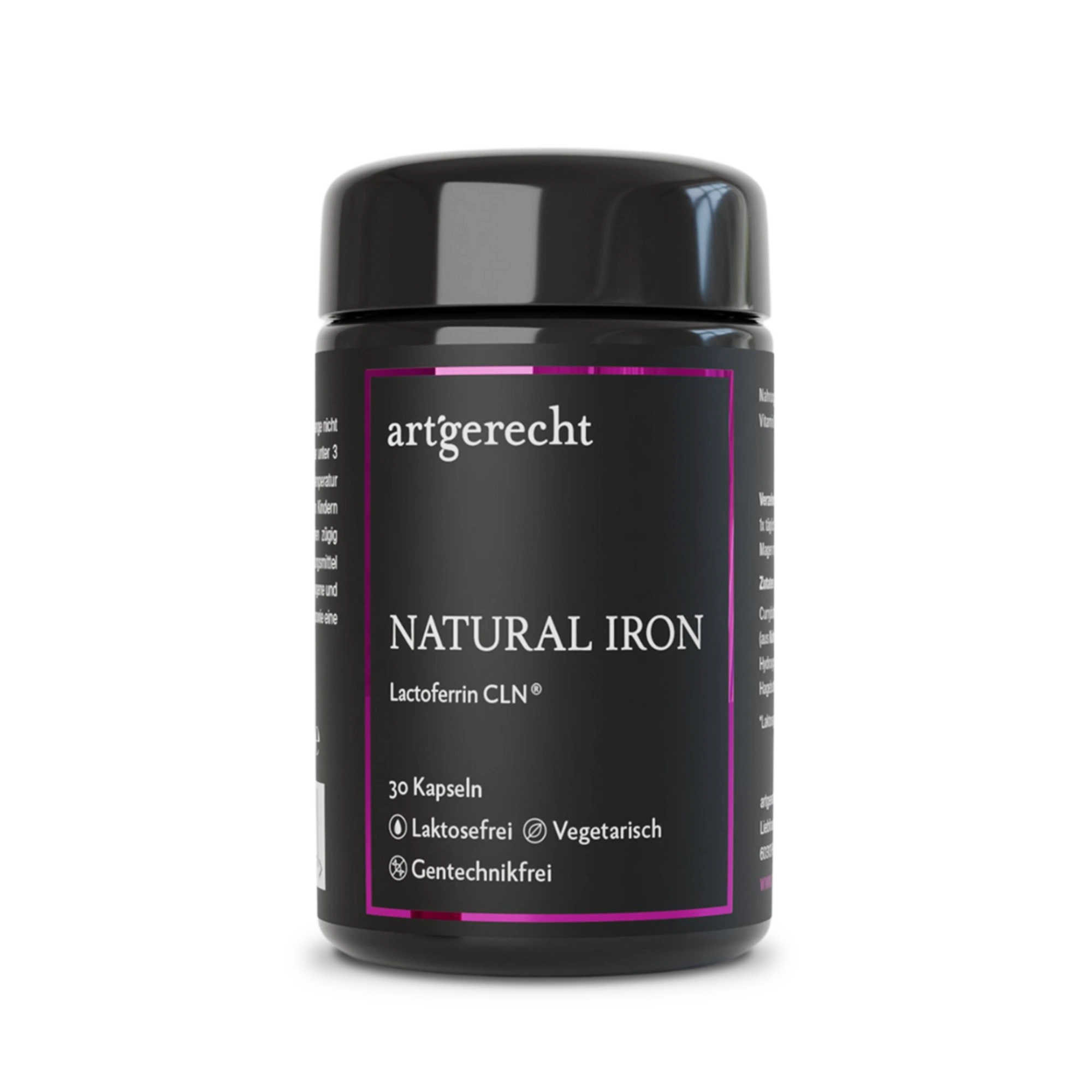
Natural Iron
For your iron balance
Specially formulated for your iron balance with plant-based curry leaf iron, Lactoferrin CLN®, and natural Vitamin C from rose hips.
30 Capsules
Regular price:
€35.90
Most read entries
3 Posts in this encyclopedia categoryMagnesiumcarbonat
Magnesiumcarbonat ist eine anorganische Magnesiumverbindung mit säure-neutralisierender (antazidischer) Wirkung. Erfahre mehr über Vorteile, Einsatzgebiete und Unterschiede zu besser resorbierbaren Magnesiumformen.
Calorie content
The choice of food influences our energy intake. The calorie content varies greatly - some provide little energy, others a lot.
Cologne list
The Cologne List ® is an important initiative in competitive sports that offers athletes guidance in the selection of nutritional supplements.