
-
 DE
DE



Collagen Structure Analysis – Methods and Significance
Collagen structure analysis examines the composition and quality of collagen fibers in tissue. It is used to diagnose connective tissue disorders and assess skin health and wound healing.
Add as Preferred SourceThings worth knowing about "Collagen Structure Analysis"
Collagen structure analysis examines the composition and quality of collagen fibers in tissue. It is used to diagnose connective tissue disorders and assess skin health and wound healing.
What is Collagen Structure Analysis?
Collagen structure analysis is a diagnostic procedure that examines the architecture, composition, and quality of collagen fibers in human tissue. Collagen is the most abundant structural protein in the human body and a key component of skin, bone, cartilage, tendons, ligaments, and blood vessels. Analyzing its structure provides essential information about the condition of connective tissue and can reveal signs of disease or aging.
Basics of Collagen
Collagen consists of long protein chains that assemble into a characteristic triple helix structure, which gives tissues their tensile strength and elasticity. To date, more than 28 collagen types have been identified, with Types I, II, and III being the most prevalent in the human body:
- Type I: Main component of skin, bone, tendons, and scar tissue
- Type II: Primary component of cartilage
- Type III: Found in blood vessels, skin, and internal organs
Alterations in collagen structure can lead to a loss of tissue integrity and function.
Methods of Collagen Structure Analysis
Several scientific and clinical methods are available for examining collagen structure:
Histological Techniques
In histology, thin tissue sections are examined under a microscope. Special staining methods such as Masson trichrome staining or Sirius Red staining visualize collagen fibers and allow assessment of their arrangement and density.
Immunohistochemistry
Immunohistochemical methods use labeled antibodies to identify and quantify specific collagen types in tissue sections. This approach enables precise differentiation between the various collagen subtypes present in a given tissue.
Electron Microscopy
Electron microscopy allows visualization of collagen structure at the ultrastructural level, revealing individual collagen fibrils and their characteristic periodic banding pattern (D-banding).
Spectroscopic Methods
Modern techniques such as Raman spectroscopy and Fourier-transform infrared spectroscopy (FTIR) analyze the molecular composition and crosslinking degree of collagen without requiring extensive tissue preparation, enabling precise characterization of protein structure.
Imaging Techniques
Non-invasive approaches including confocal reflectance microscopy, optical coherence tomography (OCT), and multiphoton microscopy (SHG imaging) allow visualization of collagen structures directly in living tissue without the need for a biopsy.
Clinical Applications
Collagen structure analysis is applied across multiple medical specialties:
- Dermatology: Assessment of skin aging, scar formation, wound healing, and conditions such as scleroderma or Ehlers-Danlos syndrome
- Orthopedics and Rheumatology: Diagnosis of cartilage damage, osteoarthritis, and collagenopathies
- Cardiology: Examination of heart valves and vessel walls in fibrotic conditions
- Oncology: Analysis of the tumor stroma, as collagen changes influence tumor growth and progression
- Aesthetic Medicine and Anti-Aging: Evaluation of skin elasticity and collagen content to guide treatment planning
Relevance in Disease
Alterations in collagen structure play a central role in numerous diseases. In fibrosis, excessive collagen deposition in tissues leads to loss of function. In hereditary connective tissue disorders such as Ehlers-Danlos syndrome or osteogenesis imperfecta, mutations in collagen genes result in structurally defective collagen. The natural aging process is also associated with a decline in collagen production and changes in fiber architecture.
Diagnosis
The choice of analytical method depends on the clinical question being addressed. An accurate diagnosis often requires a combination of imaging techniques, laboratory tests, and biopsy-based methods. Biomarkers in blood or urine, such as procollagen propeptides or collagen crosslinks, can also provide insight into collagen metabolism and turnover.
References
- Ricard-Blum S. - The Collagen Family. Cold Spring Harbor Perspectives in Biology, 2011. doi:10.1101/cshperspect.a004978
- Frantz C., Stewart K.M., Weaver V.M. - The extracellular matrix at a glance. Journal of Cell Science, 2010. doi:10.1242/jcs.023820
- Shoulders M.D., Raines R.T. - Collagen Structure and Stability. Annual Review of Biochemistry, 2009. doi:10.1146/annurev.biochem.77.032207.120833

Building Blocks for a Healthy Life
As an integrated manufacturer, we develop & produce evidence-based, patented, and non-patented formulations. We work exclusively with plant and natural extracts according to the strictest purity & quality standards.
The combination of science & modern technology with the laws of nature creates solutions consistently tailored to humans – for the highest good: health.
Skip product gallery


Subscription available
Floral
For Healthy Oral Flora & Dental Care
Formulated lozenges with Dentalac®, lactic acid bacteria, and Lactoferrin CLN®30 lozenges
Regular price:
€22.90
Discount
Subscription available
Lactoferrin 60 Capsules

For your universal protection
As one of the most valuable proteins in the body, lactoferrin is a natural component of the immune system.
60 Capsules
Sale price:
€50.00
Regular price:
€59.90
Discount
Subscription available
Multivitamin Mineral Complex
Daily basic supply: the complete spectrum of micronutrients
Natural multivitamin complex for the immune system, energy, and central body functions
30 Capsules
Sale price:
€27.90
Regular price:
€29.90
Subscription available
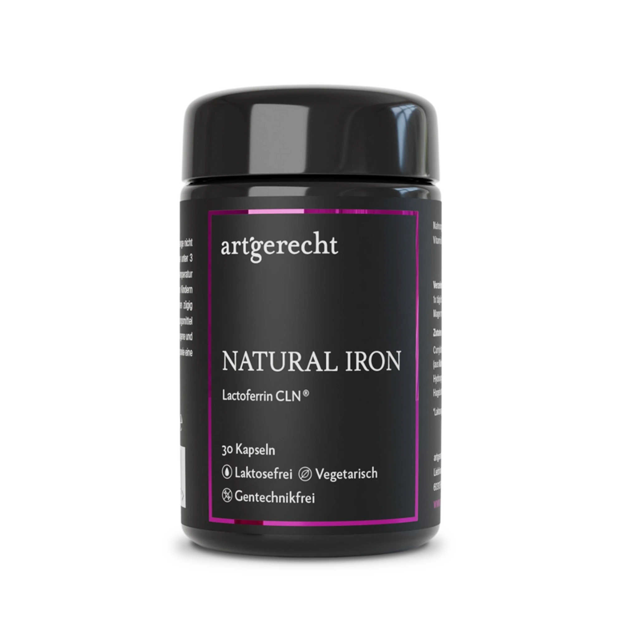
Natural Iron
For your iron balance
Specially formulated for your iron balance with plant-based curry leaf iron, Lactoferrin CLN®, and natural Vitamin C from rose hips.
30 Capsules
Regular price:
€35.90
Most read entries
3 Posts in this encyclopedia categoryMagnesiumcarbonat
Magnesiumcarbonat ist eine anorganische Magnesiumverbindung mit säure-neutralisierender (antazidischer) Wirkung. Erfahre mehr über Vorteile, Einsatzgebiete und Unterschiede zu besser resorbierbaren Magnesiumformen.
Calorie content
The choice of food influences our energy intake. The calorie content varies greatly - some provide little energy, others a lot.
Cologne list
The Cologne List ® is an important initiative in competitive sports that offers athletes guidance in the selection of nutritional supplements.