
-
 DE
DE



Bone Scintigraphy – Procedure, Uses & Findings
Bone scintigraphy is a nuclear medicine imaging technique used to visualize bone metabolism. It enables early detection of bone metastases, infections, and fractures.
Add as Preferred SourceThings worth knowing about "Bone Scintigraphy"
Bone scintigraphy is a nuclear medicine imaging technique used to visualize bone metabolism. It enables early detection of bone metastases, infections, and fractures.
What is Bone Scintigraphy?
Bone scintigraphy (also called a bone scan or skeletal scintigraphy) is a nuclear medicine imaging procedure that visualizes metabolic activity in bone tissue. Unlike conventional X-rays, which primarily show bone structure, bone scintigraphy detects functional changes in bone – often before any structural abnormalities become visible on X-ray. It is one of the most sensitive tools available for the early detection of a wide range of bone disorders.
How Does Bone Scintigraphy Work?
A small amount of a weakly radioactive substance – called a radiopharmaceutical – is injected into a vein. The most commonly used agent is Technetium-99m-labeled diphosphonate (e.g., Tc-99m-MDP). This tracer accumulates in areas of increased bone turnover, such as sites affected by tumors, inflammation, fracture healing, or metastatic disease.
After a waiting period of approximately two to four hours – allowing the tracer to distribute throughout the body – the patient is scanned using a specialized camera called a gamma camera. This device detects the gamma radiation emitted by the tracer and generates an image of metabolic activity across the entire skeleton. Areas of elevated activity appear as hot spots, while areas of decreased activity appear as cold spots.
When is Bone Scintigraphy Used?
Bone scintigraphy is indicated in a variety of clinical scenarios:
- Bone metastases: Detecting secondary tumors from known cancers such as breast, prostate, or lung cancer
- Primary bone tumors: Assessing extent and metabolic activity
- Osteomyelitis: Diagnosing bacterial bone infections
- Stress fractures and occult fractures: Identifying fractures not yet visible on X-ray
- Paget disease of bone: Evaluating the extent of abnormal bone remodeling
- Arthritis and joint disease: Assessing inflammatory activity in joints
- Avascular necrosis: Early detection of bone blood supply disruption
Procedure Overview
The examination typically takes place in several stages:
- Injection: The radiopharmaceutical is administered intravenously.
- Waiting period: Over two to four hours, the tracer distributes throughout the body. Patients are encouraged to drink plenty of fluids to promote the excretion of unbound tracer via the kidneys.
- Imaging: The scan itself usually takes 30 to 60 minutes. The patient lies still on an examination table while the gamma camera moves slowly over the entire body.
- Reporting: Images are analyzed by a nuclear medicine specialist and the findings are communicated to the referring physician.
Radiation Exposure and Safety
The radiation dose involved in bone scintigraphy is relatively low. The effective dose is approximately 3 to 6 millisieverts (mSv), depending on the radiopharmaceutical used and the patient's body weight – comparable to the natural background radiation exposure in central Europe over the course of one year. Technetium-99m has a short physical half-life of approximately six hours, meaning radioactivity decays rapidly.
In pregnant women, the procedure should only be performed when there is an urgent medical indication. Breastfeeding mothers are advised to interrupt breastfeeding for at least 24 hours following the injection.
Advantages and Limitations Compared to Other Imaging Methods
Bone scintigraphy offers several important advantages over other imaging modalities:
- High sensitivity for bone changes, often detectable earlier than on X-ray
- Whole-body survey in a single examination
- Relatively cost-effective compared to whole-body MRI
Potential limitations include:
- Lower specificity: Hot spots can have many different causes and often require further workup
- Limited anatomical detail compared to MRI or CT
- Low-level radiation exposure from radioactivity
References
- Bombardieri E. et al. - EANM procedure guidelines for bone scintigraphy. European Journal of Nuclear Medicine and Molecular Imaging, 2012.
- Aktolun C., Goldsmith S. J. (eds.) - Nuclear Medicine Therapy. Springer, New York, 2013.
- Society of Nuclear Medicine and Molecular Imaging (SNMMI) - Procedure Standard for Bone Scintigraphy. www.snmmi.org, accessed 2024.

Building Blocks for a Healthy Life
As an integrated manufacturer, we develop & produce evidence-based, patented, and non-patented formulations. We work exclusively with plant and natural extracts according to the strictest purity & quality standards.
The combination of science & modern technology with the laws of nature creates solutions consistently tailored to humans – for the highest good: health.
Skip product gallery


Subscription available
Floral
For Healthy Oral Flora & Dental Care
Formulated lozenges with Dentalac®, lactic acid bacteria, and Lactoferrin CLN®30 lozenges
Regular price:
€22.90
Discount
Subscription available
Lactoferrin 60 Capsules

For your universal protection
As one of the most valuable proteins in the body, lactoferrin is a natural component of the immune system.
60 Capsules
Sale price:
€50.00
Regular price:
€59.90
Discount
Subscription available
Multivitamin Mineral Complex
Daily basic supply: the complete spectrum of micronutrients
Natural multivitamin complex for the immune system, energy, and central body functions
30 Capsules
Sale price:
€27.90
Regular price:
€29.90
Subscription available
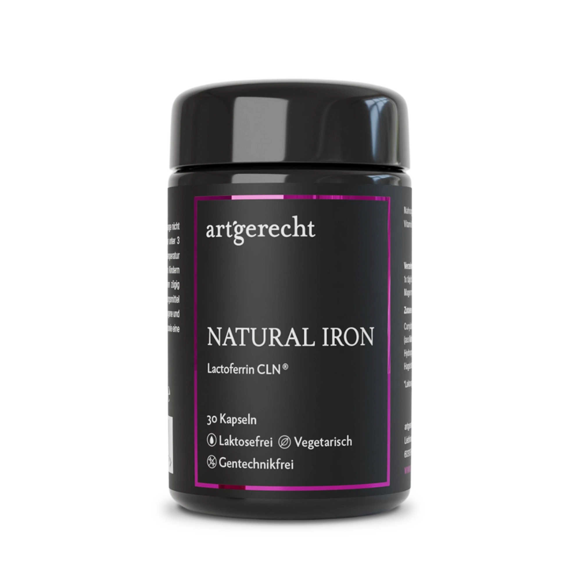
Natural Iron
For your iron balance
Specially formulated for your iron balance with plant-based curry leaf iron, Lactoferrin CLN®, and natural Vitamin C from rose hips.
30 Capsules
Regular price:
€35.90
Most read entries
3 Posts in this encyclopedia categoryMagnesiumcarbonat
Magnesiumcarbonat ist eine anorganische Magnesiumverbindung mit säure-neutralisierender (antazidischer) Wirkung. Erfahre mehr über Vorteile, Einsatzgebiete und Unterschiede zu besser resorbierbaren Magnesiumformen.
Calorie content
The choice of food influences our energy intake. The calorie content varies greatly - some provide little energy, others a lot.
Cologne list
The Cologne List ® is an important initiative in competitive sports that offers athletes guidance in the selection of nutritional supplements.