
-
 DE
DE



Cavernoma: Causes, Symptoms & Treatment
A cavernoma is a benign vascular malformation in the brain or spinal cord made up of abnormally enlarged blood vessels. It can cause bleeding, epilepsy, or neurological deficits.
Add as Preferred SourceThings worth knowing about "Cavernoma"
A cavernoma is a benign vascular malformation in the brain or spinal cord made up of abnormally enlarged blood vessels. It can cause bleeding, epilepsy, or neurological deficits.
What is a Cavernoma?
A cavernoma (also known as a cavernous malformation or cavernous hemangioma) is a benign vascular malformation consisting of a cluster of abnormally enlarged, thin-walled blood vessels. These vessels are filled with blood and surrounded by connective tissue. Cavernomas can occur anywhere in the central nervous system but are most commonly found in the brain and spinal cord. They belong to the group of vascular malformations and are not malignant tumors.
Causes
Cavernomas can develop either sporadically (without a known cause) or in a familial (hereditary) form. The familial form is associated with mutations in specific genes, particularly CCM1 (KRIT1), CCM2, and CCM3. These mutations are inherited in an autosomal dominant pattern, meaning a single mutated copy of the gene is sufficient to cause the condition. In rare cases, cavernomas may develop following radiation therapy to the brain.
- Sporadic development (most common form)
- Genetic mutation (familial cavernomatosis)
- Radiation-induced cavernomas following radiotherapy
Symptoms
Many cavernomas are completely asymptomatic and are discovered incidentally during brain imaging. When symptoms do occur, they depend strongly on the location of the cavernoma within the brain or spinal cord.
- Epileptic seizures: The most common symptom for cavernomas in the cerebral hemispheres
- Headaches: Particularly associated with acute bleeding episodes
- Neurological deficits: Visual disturbances, speech problems, paralysis, or sensory loss depending on the affected brain region
- Dizziness and balance problems: When cavernomas are located in the cerebellum or brainstem
- Spinal cord symptoms: Weakness or numbness in the limbs in cases of spinal cavernomas
Diagnosis
The most reliable method for diagnosing a cavernoma is magnetic resonance imaging (MRI). On MRI, cavernomas show a characteristic appearance often described as a "popcorn" pattern, caused by repeated small bleeds and blood breakdown products. Computed tomography (CT) is less sensitive but may reveal calcifications. Conventional angiography is generally not informative, as cavernomas lack a direct arterial blood supply.
- MRI of the brain or spine (method of choice)
- CT scan (complementary)
- Genetic testing if a familial form is suspected
- EEG in cases of epileptic seizures
Treatment
Treatment of a cavernoma depends on its location, size, associated symptoms, and bleeding risk. Asymptomatic cavernomas are typically managed conservatively with regular monitoring (a watch-and-wait approach).
Surgical Treatment
For symptomatic cavernomas, especially those with recurrent bleeding or drug-resistant epilepsy, neurosurgical removal may be recommended. The goal is complete resection of the cavernoma. Success rates are high when the lesion is surgically accessible.
Radiation Treatment
Radiation therapy (e.g., radiosurgery using Gamma Knife) is an option for cavernomas located in surgically inaccessible areas of the brain. However, its effectiveness for cavernomas is more debated than for other vascular malformations.
Medical Therapy
There is no specific drug treatment that targets the cavernoma itself. However, antiepileptic medications can be used to manage seizures associated with the condition.
Prognosis
The prognosis largely depends on the location and clinical course of the cavernoma. Many patients live without significant limitations. The annual bleeding risk is approximately 0.5 to 3 percent per year per lesion and is higher following a previous bleed. Cavernomas located in the brainstem carry a particularly elevated risk.
References
- Flemming KD, Link MJ, Christianson TJ, Brown RD Jr. - Prospective hemorrhage risk of intracerebral cavernous malformations. Neurology, 2012.
- Akers A, Al-Shahi Salman R, et al. - Synopsis of Guidelines for the Clinical Management of Cerebral Cavernous Malformations. Neurosurgery, 2017.
- Rigamonti D (ed.) - Cavernous Malformations of the Nervous System. Cambridge University Press, 2011.

Building Blocks for a Healthy Life
As an integrated manufacturer, we develop & produce evidence-based, patented, and non-patented formulations. We work exclusively with plant and natural extracts according to the strictest purity & quality standards.
The combination of science & modern technology with the laws of nature creates solutions consistently tailored to humans – for the highest good: health.
Skip product gallery


Subscription available
Floral
For Healthy Oral Flora & Dental Care
Formulated lozenges with Dentalac®, lactic acid bacteria, and Lactoferrin CLN®30 lozenges
Regular price:
€22.90
Discount
Subscription available
Lactoferrin 60 Capsules

For your universal protection
As one of the most valuable proteins in the body, lactoferrin is a natural component of the immune system.
60 Capsules
Sale price:
€50.00
Regular price:
€59.90
Discount
Subscription available
Multivitamin Mineral Complex
Daily basic supply: the complete spectrum of micronutrients
Natural multivitamin complex for the immune system, energy, and central body functions
30 Capsules
Sale price:
€27.90
Regular price:
€29.90
Subscription available
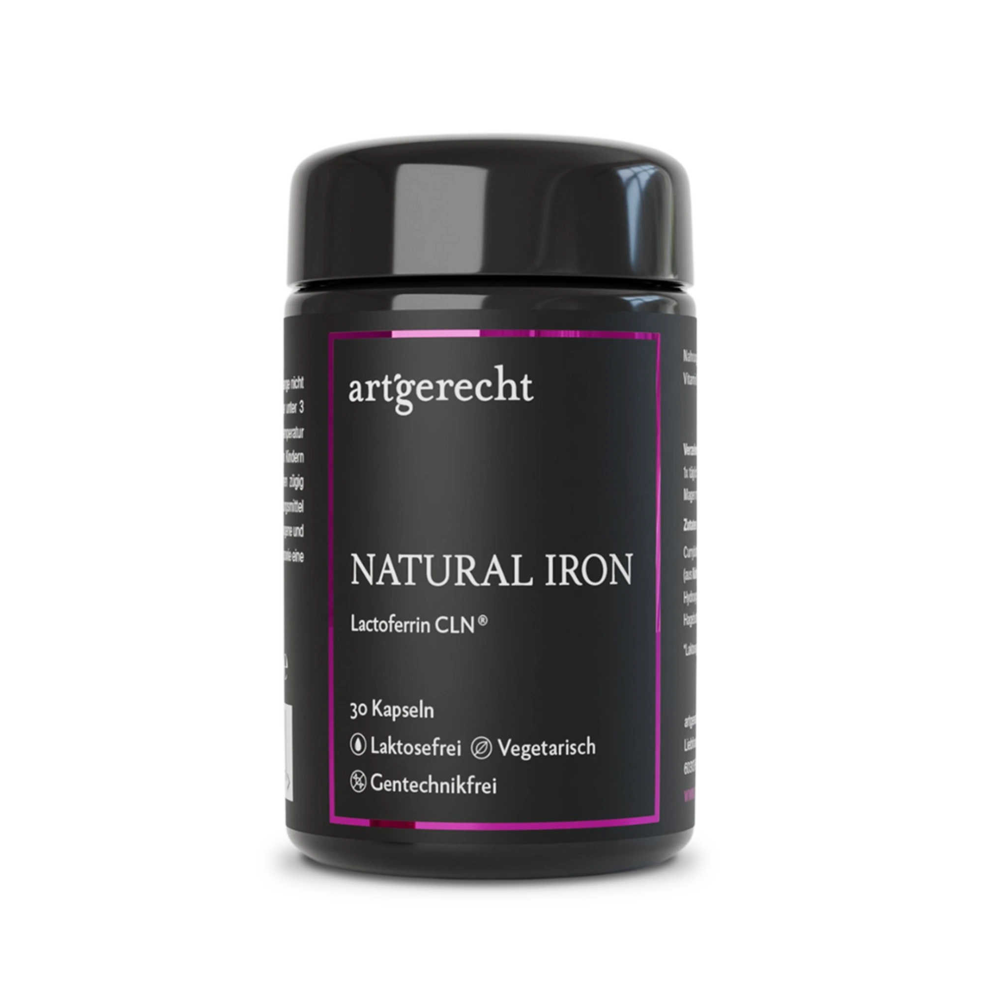
Natural Iron
For your iron balance
Specially formulated for your iron balance with plant-based curry leaf iron, Lactoferrin CLN®, and natural Vitamin C from rose hips.
30 Capsules
Regular price:
€35.90
Most read entries
3 Posts in this encyclopedia categoryMagnesiumcarbonat
Magnesiumcarbonat ist eine anorganische Magnesiumverbindung mit säure-neutralisierender (antazidischer) Wirkung. Erfahre mehr über Vorteile, Einsatzgebiete und Unterschiede zu besser resorbierbaren Magnesiumformen.
Calorie content
The choice of food influences our energy intake. The calorie content varies greatly - some provide little energy, others a lot.
Cologne list
The Cologne List ® is an important initiative in competitive sports that offers athletes guidance in the selection of nutritional supplements.