
-
 DE
DE



Fossa Hyaloidea – Anatomy and Clinical Relevance
The fossa hyaloidea is a shallow, cup-shaped depression on the front surface of the vitreous body of the eye, in which the crystalline lens rests.
Add as Preferred SourceThings worth knowing about "Fossa hyaloidea"
The fossa hyaloidea is a shallow, cup-shaped depression on the front surface of the vitreous body of the eye, in which the crystalline lens rests.
Definition
The fossa hyaloidea (also referred to as the fossa patellaris vitrei) is an anatomical structure of the human eye. It is a shallow, concave, cup-shaped depression located on the anterior surface of the vitreous body (corpus vitreum), into which the posterior surface of the crystalline lens fits. This depression provides stable support for the lens and is an essential component of the anatomical architecture of the anterior segment of the eye.
Anatomy and Location
The vitreous body occupies the majority of the interior of the eye and consists of approximately 99% water, along with a scaffold of collagen fibers and hyaluronic acid. On its anterior surface, the vitreous body features the fossa hyaloidea – a circular, concave indentation that precisely corresponds to the convex posterior surface of the crystalline lens, allowing the lens to rest securely within it.
At the center of the fossa hyaloidea lies the anterior opening of the hyaloid canal (also known as Cloquet canal), an embryonic remnant of the former vascular system within the vitreous, which regresses during fetal development.
Functional Significance
The fossa hyaloidea serves several important functions within the eye:
- Stabilization of the crystalline lens: The depression holds the lens in its correct position, supported by the zonular fibers (zonula ciliaris), which suspend the lens from the ciliary body.
- Mechanical cushioning: The vitreous body absorbs shocks and protects both the lens and the retina from mechanical injury.
- Optical function: The precise positioning of the lens within the fossa hyaloidea is essential for accurate focusing of light onto the retina, enabling sharp vision.
Clinical Relevance
The fossa hyaloidea holds particular clinical significance in the context of ophthalmic surgery and ocular disease:
Cataract Surgery
During the surgical removal of a clouded crystalline lens (cataract) and implantation of an intraocular lens (IOL), the integrity of the fossa hyaloidea and the adjacent vitreous body is critically important. Damage to this region can lead to complications such as vitreous prolapse, where vitreous tissue enters the anterior chamber of the eye.
Vitreous Detachment and Degeneration
With increasing age, posterior vitreous detachment may occur, in which the vitreous body separates from the retina. Structural changes in the vitreous indirectly affect the anatomy of the fossa hyaloidea region as well.
Intraocular Surgery
During vitrectomy procedures (surgical removal of the vitreous body), the surgeon must have a thorough understanding of the anatomical relationship between the fossa hyaloidea, the lens, and the ciliary body in order to avoid inadvertent injury.
Embryology
During embryonic eye development, the hyaloid artery courses through the vitreous body to supply the developing lens. After birth, this vessel regresses and leaves behind the hyaloid canal, whose anterior opening is located within the fossa hyaloidea. If the vessel fails to regress fully after birth, the condition is referred to as a persistent hyaloid canal or persistent fetal vasculature (PFV), which may cause visual impairment.
References
- Forrester, J. V., Dick, A. D., McMenamin, P. G., Roberts, F. & Pearlman, E. (2016). The Eye – Basic Sciences in Practice. 4th ed. Elsevier Saunders.
- Remington, L. A. (2012). Clinical Anatomy and Physiology of the Visual System. 3rd ed. Butterworth-Heinemann.
- Standring, S. (ed.) (2020). Gray's Anatomy – The Anatomical Basis of Clinical Practice. 42nd ed. Elsevier.

Building Blocks for a Healthy Life
As an integrated manufacturer, we develop & produce evidence-based, patented, and non-patented formulations. We work exclusively with plant and natural extracts according to the strictest purity & quality standards.
The combination of science & modern technology with the laws of nature creates solutions consistently tailored to humans – for the highest good: health.
Skip product gallery


Subscription available
Floral
For Healthy Oral Flora & Dental Care
Formulated lozenges with Dentalac®, lactic acid bacteria, and Lactoferrin CLN®30 lozenges
Regular price:
€22.90
Discount
Subscription available
Lactoferrin 60 Capsules

For your universal protection
As one of the most valuable proteins in the body, lactoferrin is a natural component of the immune system.
60 Capsules
Sale price:
€50.00
Regular price:
€59.90
Discount
Subscription available
Multivitamin Mineral Complex
Daily basic supply: the complete spectrum of micronutrients
Natural multivitamin complex for the immune system, energy, and central body functions
30 Capsules
Sale price:
€27.90
Regular price:
€29.90
Subscription available
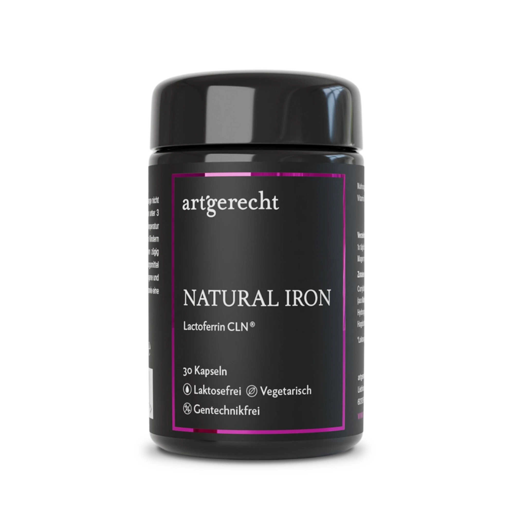
Natural Iron
For your iron balance
Specially formulated for your iron balance with plant-based curry leaf iron, Lactoferrin CLN®, and natural Vitamin C from rose hips.
30 Capsules
Regular price:
€35.90
Most read entries
3 Posts in this encyclopedia categoryMagnesiumcarbonat
Magnesiumcarbonat ist eine anorganische Magnesiumverbindung mit säure-neutralisierender (antazidischer) Wirkung. Erfahre mehr über Vorteile, Einsatzgebiete und Unterschiede zu besser resorbierbaren Magnesiumformen.
Calorie content
The choice of food influences our energy intake. The calorie content varies greatly - some provide little energy, others a lot.
Cologne list
The Cologne List ® is an important initiative in competitive sports that offers athletes guidance in the selection of nutritional supplements.