
-
 DE
DE



Intestinal Villus Regeneration – Causes and Support
Intestinal villus regeneration is the process by which the small intestinal lining repairs and rebuilds its finger-like projections that are essential for nutrient absorption.
Add as Preferred SourceThings worth knowing about "Intestinal Villus Regeneration"
Intestinal villus regeneration is the process by which the small intestinal lining repairs and rebuilds its finger-like projections that are essential for nutrient absorption.
What is Intestinal Villus Regeneration?
Intestinal villus regeneration refers to the biological process by which the small fingerlike projections of the small intestinal lining – known as intestinal villi (Villi intestinales) – are repaired and restored following damage. These villi are essential for the absorption of nutrients, vitamins, and minerals from digested food. Impaired or incomplete regeneration can lead to significant nutritional deficiencies and digestive disorders.
Structure and Function of Intestinal Villi
Intestinal villi are microscopic projections of the small intestinal mucosa that, together with the microvilli (brush border), massively increase the absorptive surface area of the small intestine. In a healthy adult, this mechanism creates a total surface area of approximately 200–300 square metres. The villi are responsible for absorbing:
- Carbohydrates (e.g. glucose, fructose)
- Proteins (as amino acids and peptides)
- Fats and fat-soluble vitamins (A, D, E, K)
- Water-soluble vitamins (e.g. B12, folate)
- Minerals and trace elements (e.g. iron, calcium, zinc)
Causes of Villous Damage
Various diseases and factors can damage the intestinal villi, triggering the need for regeneration:
- Coeliac disease: An autoimmune condition in which gluten triggers an immune response leading to villous atrophy (flattening and loss of villi).
- Crohn disease: A chronic inflammatory bowel disease that can affect all layers of the intestinal wall.
- Infectious enteritis: Bacterial or viral intestinal infections (e.g. rotavirus, Salmonella) can temporarily damage the mucosal lining.
- Food allergies: Certain allergens can provoke local inflammatory reactions and mucosal injury.
- Chemotherapy and radiation: Oncological treatments can damage the rapidly dividing cells of the intestinal mucosa.
- Parasitic infections: Pathogens such as Giardia lamblia can impair villous architecture.
- Malnutrition and nutrient deficiency: Deficiencies in zinc, vitamin A, and glutamine in particular can inhibit mucosal renewal.
Mechanism of Intestinal Villus Regeneration
The intestinal mucosa has a remarkable capacity for self-renewal. Regeneration is driven by stem cells located in the so-called crypts of Lieberkuehn – deep invaginations between the villi. These stem cells continuously divide and migrate upward along the villus, differentiating into specialised cells, primarily enterocytes (absorptive cells). In a healthy individual, the complete renewal cycle of the intestinal mucosa takes approximately 3–5 days.
In the context of damage – for example, from coeliac disease or infection – this process is disrupted. Once the underlying cause is removed (e.g. a gluten-free diet in coeliac disease), regeneration can begin, but depending on the severity of injury and individual factors, full recovery may take months to years.
Factors Influencing Regeneration
Several factors affect the speed and quality of intestinal villus regeneration:
- Diet: A balanced, nutrient-rich diet supports mucosal renewal. Specific nutrients such as glutamine, zinc, vitamin A, and short-chain fatty acids (e.g. butyrate) play a particularly important role.
- Probiotics and gut microbiota: A healthy gut microbiome promotes mucosal integrity and supports regeneration.
- Age: Cell division and regenerative capacity slow with increasing age.
- Inflammation control: Active inflammation inhibits regeneration; treating the underlying condition is therefore essential.
- Medications: Certain drugs (e.g. non-steroidal anti-inflammatory drugs) can place additional stress on the mucosa.
Diagnosis and Monitoring
Assessment of intestinal villus regeneration is typically performed via small intestinal biopsy during upper endoscopy (gastroscopy), with tissue samples taken from the duodenum or proximal small intestine. Histologically, the degree of villous atrophy is graded using the Marsh classification. Complementary blood tests for nutritional deficiencies and antibody testing (e.g. in coeliac disease) may also be used.
Therapeutic Approaches to Support Regeneration
The most important measure is eliminating the underlying cause. In addition, the following strategies are commonly applied:
- Gluten-free diet in coeliac disease – the cornerstone of treatment
- Nutrient supplementation to correct deficiencies (iron, folate, vitamin D, B12, zinc)
- Enteral or parenteral nutrition in cases of severe mucosal damage
- Probiotics to support gut flora balance
- Anti-inflammatory therapy for chronic inflammatory conditions
- Mucosal-protective substances such as L-glutamine or zinc carnosine
References
- Oberhuber G. et al. - The histopathology of coeliac disease: time for a standardized report scheme for pathologists. European Journal of Gastroenterology and Hepatology, 1999; 11(10): 1185–1194.
- Marsh MN. - Gluten, major histocompatibility complex, and the small intestine. Gastroenterology, 1992; 102(1): 330–354.
- World Gastroenterology Organisation (WGO) - Global Guidelines: Celiac Disease. WGO, 2016. Available at: https://www.worldgastroenterology.org

Building Blocks for a Healthy Life
As an integrated manufacturer, we develop & produce evidence-based, patented, and non-patented formulations. We work exclusively with plant and natural extracts according to the strictest purity & quality standards.
The combination of science & modern technology with the laws of nature creates solutions consistently tailored to humans – for the highest good: health.
Skip product gallery


Subscription available
Floral
For Healthy Oral Flora & Dental Care
Formulated lozenges with Dentalac®, lactic acid bacteria, and Lactoferrin CLN®30 lozenges
Regular price:
€22.90
Discount
Subscription available
Lactoferrin 60 Capsules

For your universal protection
As one of the most valuable proteins in the body, lactoferrin is a natural component of the immune system.
60 Capsules
Sale price:
€50.00
Regular price:
€59.90
Discount
Subscription available
Multivitamin Mineral Complex
Daily basic supply: the complete spectrum of micronutrients
Natural multivitamin complex for the immune system, energy, and central body functions
30 Capsules
Sale price:
€27.90
Regular price:
€29.90
Subscription available
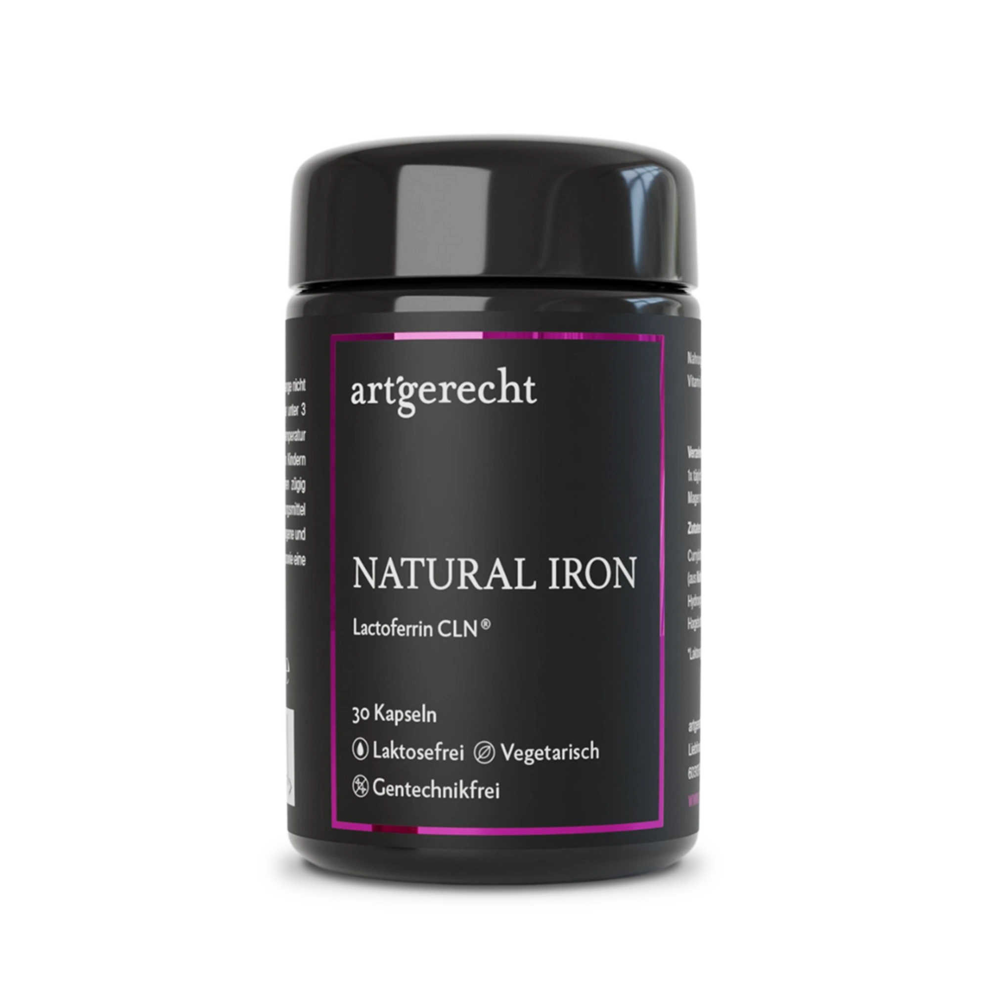
Natural Iron
For your iron balance
Specially formulated for your iron balance with plant-based curry leaf iron, Lactoferrin CLN®, and natural Vitamin C from rose hips.
30 Capsules
Regular price:
€35.90
Most read entries
3 Posts in this encyclopedia categoryMagnesiumcarbonat
Magnesiumcarbonat ist eine anorganische Magnesiumverbindung mit säure-neutralisierender (antazidischer) Wirkung. Erfahre mehr über Vorteile, Einsatzgebiete und Unterschiede zu besser resorbierbaren Magnesiumformen.
Calorie content
The choice of food influences our energy intake. The calorie content varies greatly - some provide little energy, others a lot.
Cologne list
The Cologne List ® is an important initiative in competitive sports that offers athletes guidance in the selection of nutritional supplements.